

ZeroCarbonPave: Bituminous pavements with biochar to reduce the carbon footprint of roads
July 14, 2026
ArchiSpace: Sustainable architecture to design future human habitats on the Moon and Mars
September 1, 202620/07/2026
A team from the Matrix Analysis and Discrete Potential Theory (MAPTHE) research group at the Universitat Politècnica de Catalunya - BarcelonaTech (UPC), and the Department of Mathematics at CUNEF Universidad in Madrid, have participated in the development of MARIA, a prototype of a portable, flexible and radiation-free device designed to improve the early detection of breast cancer. The device can identify tumours through electrical conductivity, with the aim of enabling its future use in primary care settings.
In 2025, approximately 37,600 women were diagnosed with breast cancer in Spain, according to data published by the Spanish Society of Medical Oncology (SEOM). It is the most common type of cancer among women in Spain: an estimated one in eight women will develop breast cancer during their lifetime. The average five-year survival rate is 85%, thanks to medical advances and, above all, screening programmes.
Against this background, a UPC research team has developed MARIA, a pioneering medical prototype designed to radically transform the early detection of breast cancer and complement traditional mammography. The device features a flexible fabric membrane that can identify tumour abnormalities using electrodes that detect electrical conductivity through an innovative algorithm. It is therefore a non-invasive, radiation-free and easy-to-use system.
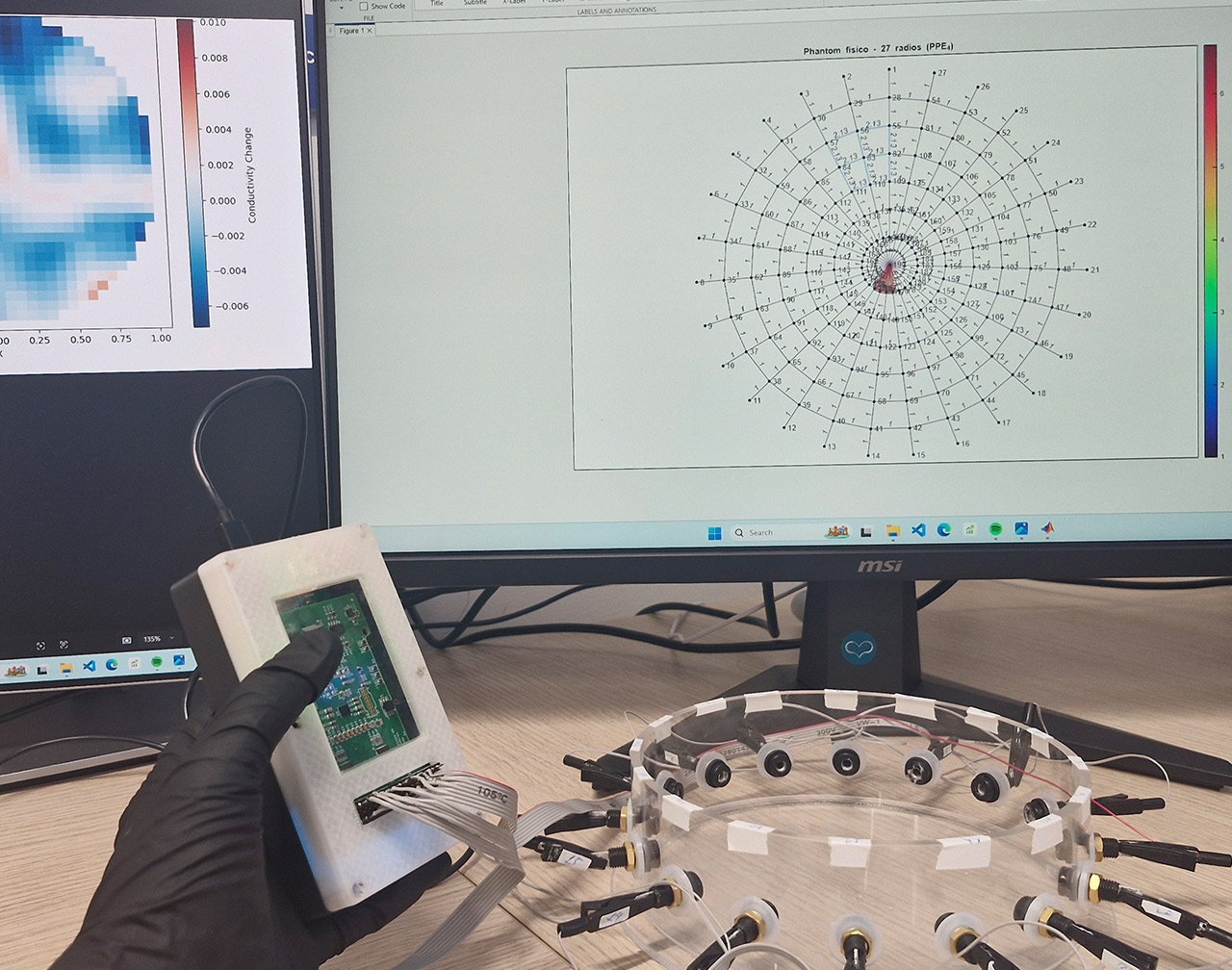
As part of the project, a new algorithm has been developed to address the inverse conductivity problem. This involves estimating the distribution of electrical conductivity inside a body using current and voltage measurements obtained solely from its surface. It is a particularly complex and unstable mathematical and numerical problem, as small errors or noise in the measurements can produce significant changes in the reconstruction.
This mathematical problem also provides the theoretical basis for Electrical Impedance Tomography (EIT), a non-invasive imaging technique with medical applications. The algorithm developed by the team and incorporated into the MARIA device reduces sensitivity to measurement noise and produces more robust tomographic reconstructions.
The scientific principle underlying the research is clear: tumours need blood to grow and, because they are more highly vascularised, their electrical conductivity is significantly higher than that of healthy tissue. By applying electrical potentials and measuring currents externally, the system can identify the location of an abnormality from the increase in conductivity.
Unlike current mammograms, which can be uncomfortable and invasive because they require breast compression, this wearable prototype is designed to be highly user-friendly and suitable for mass production, which could reduce triage costs.
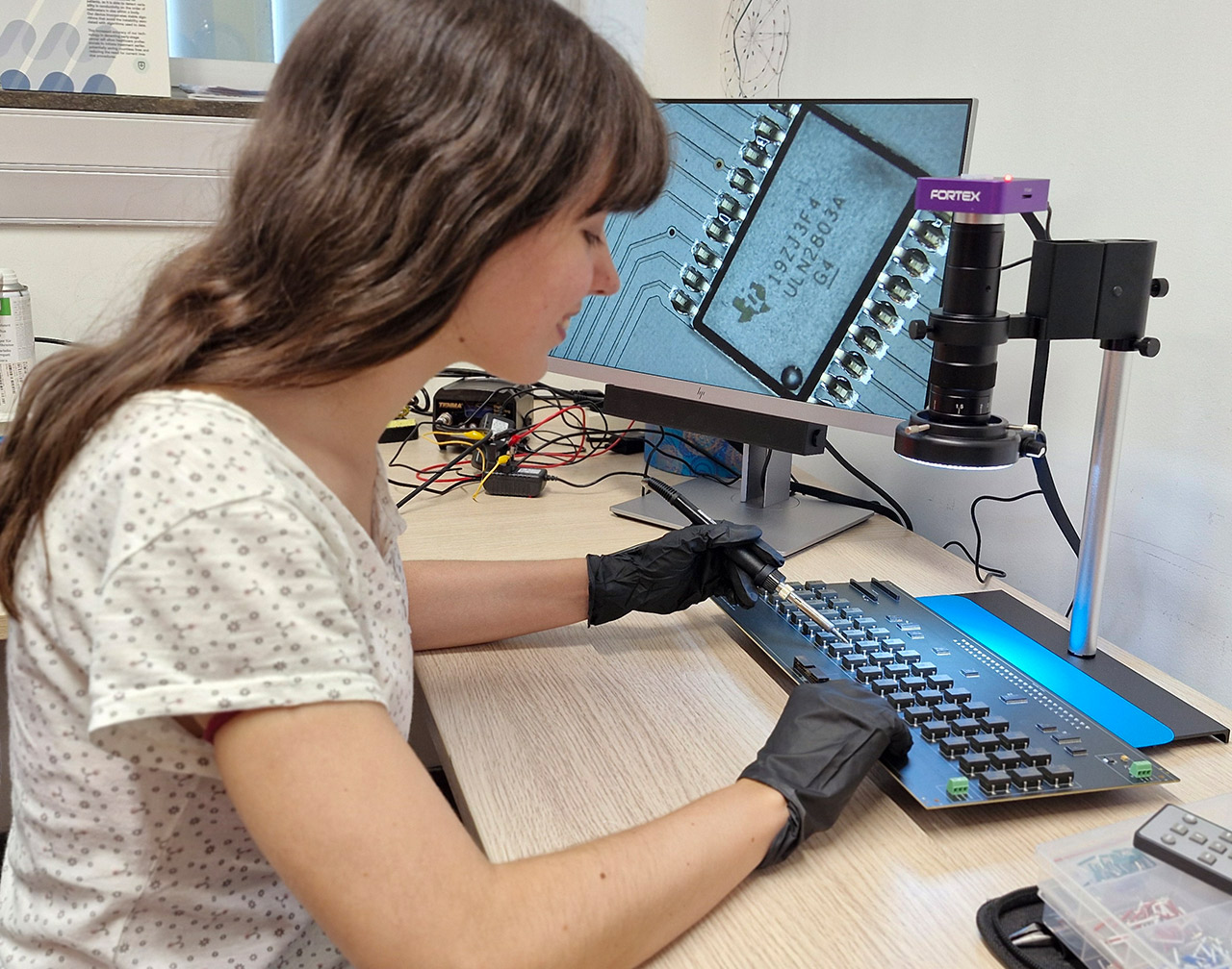
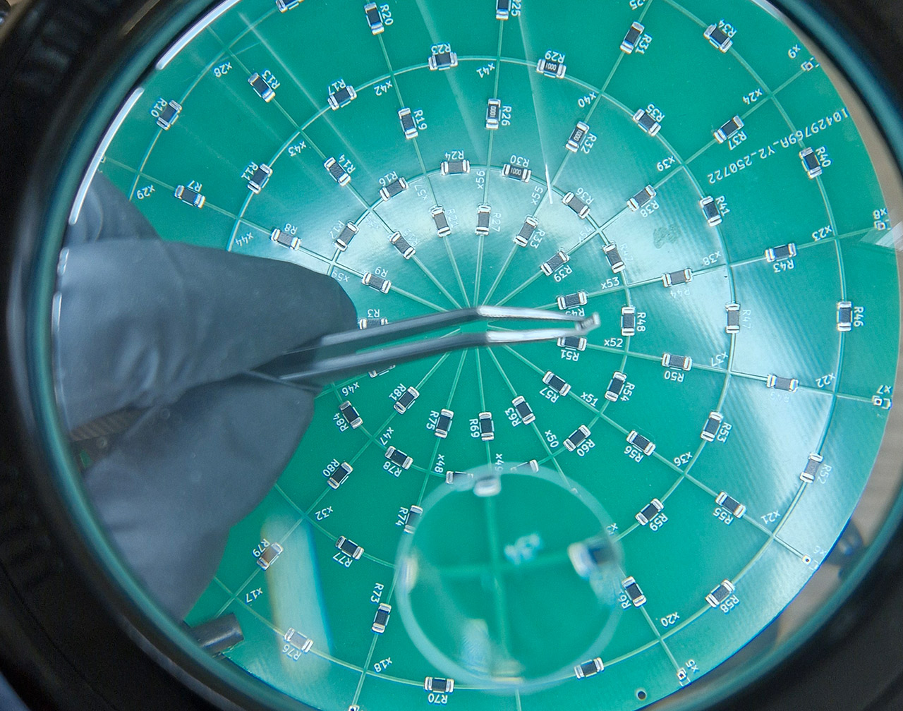
The device uses electrodes that adapt optimally to any breast shape. To achieve this, the team is working with flexible electronics to ensure good contact with the skin.
The complete absence of radiation opens the door to a range of preventive possibilities that have not previously been viable:
- Use among excluded at-risk groups: it could be used safely by pregnant women or young women aged 15 and over, for whom mammography is completely contraindicated because of cumulative radiation exposure.
- Detection in primary care: the intention is for MARIA to be used directly by GPs. Healthcare staff would place the membrane over the patient’s breast and view an almost immediate tomographic image on a screen. Should an abnormality be detected, the patient would be referred to the relevant hospital service.
- Other medical applications, such as brain monitoring in newborn babies.
Budget and Funding
A patent application covering the construction of the device’s membrane has already been submitted. The system is currently at the preclinical stage: the algorithms and electronics have been validated using breast phantoms made from agar gel and salt, which simulate the density and conductivity of breast tissue. The next planned steps include ex vivo testing using animal tissue collected post-mortem, followed by further trials in collaboration with the Germans Trias i Pujol Research Institute.
The project has received €150,000 in funding from the Agència de Gestió d’Ajuts Universitaris i de Recerca (AGAUR) under the Generalitat de Catalunya’s 2024 Indústria del Coneixement funding scheme. It will run for one year and nine months (December 2024 - September 2026).